An important step towards live imaging in proton therapy
Researchers at the Helmholtz-Zentrum Dresden-Rossendorf (HZDR) want to build the world’s first prototype that tracks moving tumors with magnetic resonance imaging (MRI) in real time during proton therapy. They are combining a rotating open MRI device, designed for the LINAC-MR system from Alberta Health Services, with an actively scanned clinical-akin proton beam at OncoRay, the Dresden-based National Center for Radiation Research in Oncology. The rotating MRI device is produced by ASG Superconductors, using their helium-free superconducting magnesium diboride (MgB2) magnet. Real-time MRI would make it possible to synchronize the proton beam to tumor motion during radiation dose delivery. So far, this has been impossible and therefore was a limiting factor for treating moving tumors with this promising cancer therapy.
Proton therapy is an emerging radiation technology to irradiate tumors while sparing the surrounding tissue better than conventional X-ray-based radiation due to the finite penetration depth of the protons. However, the effectiveness of proton therapy is limited by tumor motion, anatomical changes during therapy, and set-up inaccuracies in patient positioning. “With onboard X-ray imaging currently available at the proton therapy system, we do not obtain high-contrast live images of where we are aiming at and what we are hitting,” says Dr. Aswin Hoffmann of the HZDR Institute of Radiooncology – OncoRay, describing the fundamental problem. “Therefore, when we treat moving tumors, we have to use rather large safety margins around the tumor tissue, reducing the targeting accuracy and increasing the risk of side effects in surrounding tissues.”
Experts were skeptical
Especially with tumors that are constantly moving during respiration or digestion, the healthy tissue immediately surrounding the tumor might receive too much radiation dose, while the tumor tissue itself receives too little. Until now, in proton therapy no suitable imaging technology to visualize the tumor and its motion during radiation with high soft-tissue contrast has been available. The solution could be magnetic resonance imaging, which would offer several advantages, as Hoffmann explains: “MRI provides unsurpassed soft-tissue contrast combined with high spatial and temporal resolution. Also, unlike X-ray imaging, MRI doesn’t deposit any additional ionizing dose into the tissue.” However, integrating MRI into proton therapy systems proves to be a major technological challenge. For a long time, so-called in-beam MRI was deemed impossible.
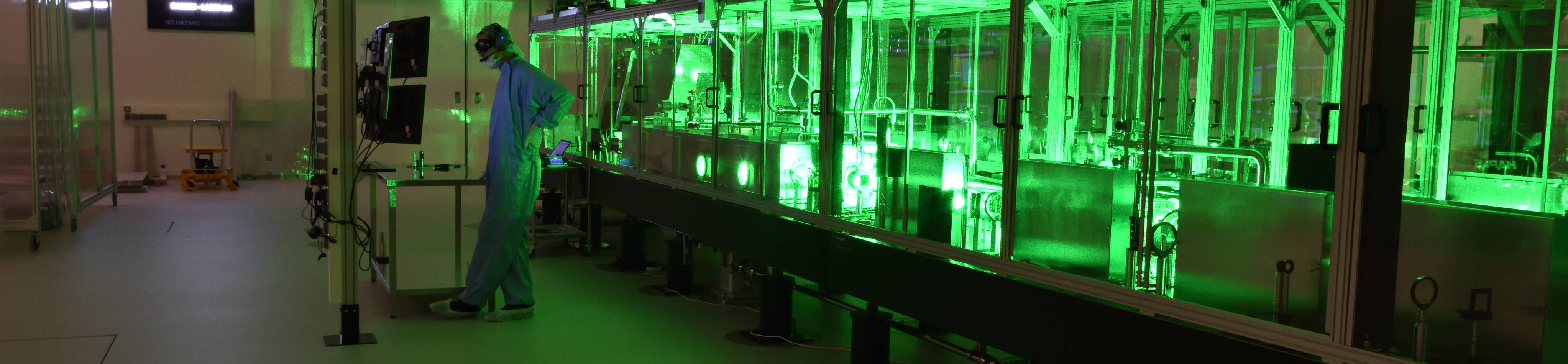
To acquire sharp and geometrically accurate images, MRI scanners operate with precisely predefined magnetic fields. At the cyclotron, the protons are accelerated by a rapidly varying (radiofrequency) electric field and kept to a spiral trajectory by a static magnetic field. During transport into the radiation room, the beam is also guided and kept in shape by magnetic fields. “Experts postulated that these electromagnetic fields would interfere with the MRI scanner and vice versa, distorting the MRI image and impacting the proton dose distribution delivered in tissue,” explains Aswin Hoffmann. In recent years, the medical physicist and his research group have been able to demonstrate, for the first time worldwide, that it is generally technologically possible to combine both systems, to experimentally confirm the presence of these effects, but also to be able to significantly compensate for them. To this end, they placed an open low-field MRI scanner with a magnetic field strength of 0.22 Tesla (MRJ2200 – ASG Superconductors Paramed MRI Unit) into the proton beam path and showed good MR image quality during irradiation with a static beam.
More information and news source: https://www.hzdr.de/db/Cms?pOid=63270&pNid=3438